


Hand and foot onychomycosis is characterized by a variety of symptoms. The appearance of nail fungus depends on the type of pathogen (there are about 50 in total), the location of the primary infection, and concomitant diseases. There are several of the most common varieties of onychomycosis, which have their own characteristics. Identification of pathology at the initial stage allows treatment only with the help of local funds. Otherwise, you will need to take systemic antifungals. With an advanced form of the disease and full damage, restoration of the nail plate becomes impossible.
Onychomycosis: a brief description
Onychomycosis is a fungal infection of the nail plates of the legs (in 80% of cases) or of the hands. At risk are older people (including 40% of people over 60 are sick), as well as those who suffer from pathologies such as:
- psoriasis;
- diabetes mellitus (prevalence three times higher than among other population groups) and other endocrine diseases;
- violation of the blood supply to the extremities as a result of cardiovascular pathologies, obliteration processes in the blood vessels leading to their narrowing; angiotrophoneurosis, varicose veins;
- oncological diseases;
- hemorrhagic sarcomatosis (multiple skin cancers);
- dermatoses associated with a violation of the keratinization process of cells, ichthyosis;
- fractures of the bones of the hand or foot;
- other serious somatic diseases, which cause a general exhaustion and a decrease in the body's defenses.
The following specialties belong to the occupational risk group:
- miners;
- Athletes;
- employees of public institutions;
- metallurgists;
- military personnel;
- medical workers;
- industrial workers;
- cooks and other workers whose activities are associated with frequent contact with water.
Among all nail diseases, onychomycosis ranks third in terms of prevalence and the total number of people carrying the disease is one fifth of the world's population. The danger of the disease lies in the fact that the focus of the fungal infection can exist for a long time and be an active source for infecting other people, including members of your own family.
In addition, fungi cause a general sensitization of the body as a result of the production of toxins, which contributes to the development of allergic and dermatological diseases in the patient. Therefore, it is important to identify the fungus at the initial stage and stop its growth, spread through the lymph and blood. Infection occurs as follows:
- in public places such as bathrooms, saunas, swimming pools, gyms;
- through shared household items (rugs, cloths, towels);
- during nail manicure treatment;
- when wearing someone else's shoes.
Risk factors are also:
- wear tight shoes;
- hot and humid climate;
- increased sweating of the legs;
- weakened immunity;
- taking hormonal and antibacterial drugs;
- Lesions and dystrophic changes in the nail plates.
Varieties of the disease.
There are several types of onychomycosis, which are classified according to various criteria. These characteristics help determine what nail fungus looks like on the hands and feet.
By type of pathogen. The defeat of the nail plate by one type of pathogen occurs in ¾ of all cases, two - in 16%, three - in 9%. The most common mixed infection occurs in elderly patients. The most common pathogens are:
- The dermatophyte fungi, the most frequent are Trichophyton rubrum (80% of all cases) and mentagrophytes var. Interdigitale (about 10% of cases). Epidermophyton floccosum, T. violaceum, and T. tonsurans are less common (3% of the total cases).
- Yeast fungi of the genus Candida, which affect more nail plates on the hands (in 40% of cases) than on the legs. The disease is usually accompanied by chronic candidiasis of the skin and mucous membranes. Several species of Candida exist as symbionts on the skin of a healthy person.
- Non-dermatophyte molds: Scytalidium dimidiatum and hyalinum, Onychocola canadensis. It is found most often in countries with hot and humid climates.
According to the shape of the nail plate injury. In the same patient, the types of damage can be combined. Depending on the transformations on the nail, the following forms are distinguished:
- Normotrophic: only a color change (yellowness at the distal free edge). The normal shape and thickness of the plate are kept for a long time. At the edges, thickenings are formed as a result of excessively rapid division of horny cells under the nails.
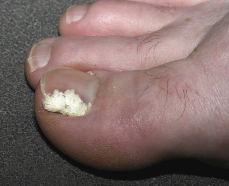
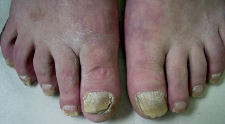
- Hypertrophic: in addition to yellowing and opacity, there is thickening and deformation of the nail plates. Over time, they acquire a transverse striation, turn dirty gray, loose at the distal edge.
- Atrophic: severe destruction of the nail occurs, the subungual skin is exposed and covered with friable and crumbling masses.
- By the type of onycholysis: there is a thinning of the nail plates, which are separated from the nail bed. The color is dull, dirty gray or yellowish; in the root area, the color may remain unchanged.
Damage location:
- Distal lateral onychomycosis.
- White surface. By scraping the damage, you can determine its superficial nature.
- Proximal subungual.
- Total dystrophic.
The first type is the most common and the origin of the lesion is infected skin. The beginning of the process occurs through the distal part - the free edge towards the root, which is why this variety received its name. In the initial stage, the nail plate retains its normal appearance, but then gradually separates from the bed and turns yellowish. In some cases, its thickening is observed. In the final stage, the color of the nail acquires different shades (from green and blue to brown), depending on the contamination of bacteria.
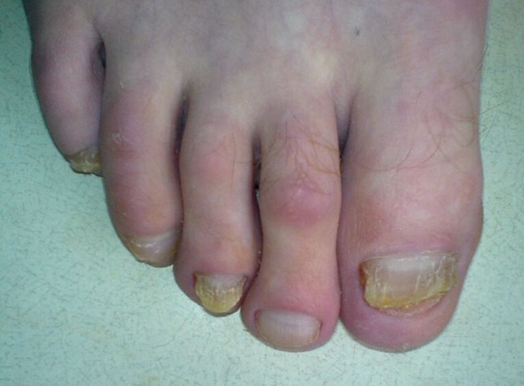
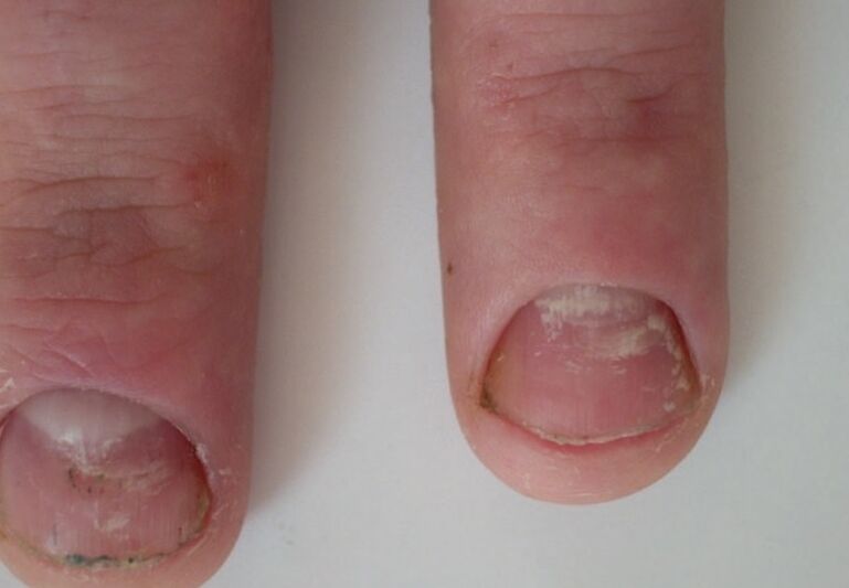
The second form of onychomycosis is characterized by the appearance of white spots, streaks that appear on the outer surface of the nail and gradually spread to the entire plate. Over time, the spots change color to yellow or yellowish-brown. In most cases, this lesion is associated with the seeding of Trichophyton mentagrophytes or molds of the genus Aspergillus. This form of the disease is more common among the elderly. The nail root and the bed, as a rule, remain intact, and the plate crumbles and turns gray or brown.
The subungual lesion is the rarest form. Its causative agents can be all three types of pathogens. The spread of the infection occurs from the skin or the lateral ridge towards the root of the nail. Spots appear in the central part of the nail plate or in the hole, and later it comes off very quickly. The nail bed and root are not inflamed, but a secondary bacterial infection often occurs, causing the nail to darken.
With the full form of the disease, the entire area of the nail is affected, which in many cases is accompanied by its complete destruction. The root disappears or thickens pathologically, as a result of which normal plaque formation is no longer possible. In the final stage of the disease, only its crumbled remains are observed. This form of onychomycosis is usually the case with candidiasis. The causative agents can also be epidermophytic fungi.
The first signs of the disease
There are symptoms that help to recognize a yeast infection in the early stage. The patient may have one or more of the following:
- loss of transparency and shine on the nail plate;
- the color of the nail becomes whitish or yellowish;
- the edge of the nail becomes uneven, thin, smooth;
- the appearance of yellow or white spots, stripes along the edges of the plate (on the hands, in the center of the nail plate);
- some types of fungus cause nail damage only on the first toe, the rest remains unchanged (these include superficial white onychomycosis, which usually damages the nail of the first toe, less often the little finger);
- the plate is moved away from the bed 1-2 mm.
For a long time, the pathological process can be localized only along the distal edge of the plate, so it is possible to stop the infection by avoiding deformation, detachment of the nail plate and damage to the root, thus which has irreversible consequences.
As the process progresses, other signs appear:
- white stripes that go from the free edge to the root;
- Nail "rib" in transverse direction;
- your weight loss;
- chipping of the plaque and other symptoms corresponding to a certain type of onychomycosis.
Superficial white onychomycosis almost never appears on the fingernails. Sometimes this form of the disease is combined with a distal one. When infected with Т. rubrum, the damage is usually multiple in nature. When infected with fungi of the genus Candida (in more rare cases, with mold infection), an inflammation of the posterior or lateral nail ridges occurs first, accompanied by the following symptoms:
- redness, compaction and swelling of the roller;
- changing its shape;
- the appearance of white scales along the edge of the roller;
- separation of the cuticle and its destruction;
- pain at the site of injury on palpation;
- in rare cases, when pressed, a small amount of pus is released.
In children, the disease in the initial stage is characterized by the following features:
- rough surface of the nail;
- the shape is often unchanged;
- defeat in most cases, at the distal edge;
- an active fungal process is also observed on the skin of the feet;
- in more rare cases, subungual hyperkeratosis is detected.
If the nail turns bright green, this indicates the addition of a secondary bacterial infection - Pseudomonas aeruginosa, and the black color indicates a Proteus infection.
Difference from other pathologies.
Nail changes, similar to onychomycosis, are also seen in other diseases:
- psoriasis (exfoliation of the nail along the edge, its thickening, irregularity "thimble" of the surface, peeling along the ridges, yellowing, destruction of the nail);
- lichen planus (deep fissure in the center of the plate, longitudinal thickenings, subungual hyperkeratosis, rupture along the distal edge, increased brittleness, loss of the nail through cleft);
- eczema (transverse grooves, softening of the tissue, peeling along the edge, thickening of the roller);
- trichophytosis of the nails (in this case, there is also a smooth or hairy skin lesion).
Since the external symptoms of onychomycosis can coincide with non-fungal diseases, a microscopic examination and seeding of the pathogen is required to make an accurate diagnosis.
Treatment
In the initial stage of the disease, if no more than half of the nail plate is affected, treatment can be carried out using only local remedies. The tactics of therapy also depend on the form of the disease.
Long-term multiple onychomycosis with bacterial complications will need to be treated for a longer time, over several months, with the help of systemic antifungal medications. Local therapy is carried out in cases where there is a high risk of side effects, in pregnant and lactating women, in people with liver disease, kidney disease, drug allergies. The disadvantage of local drugs is that they cannot penetrate the root of the nail, and if the matrix is damaged, such treatment is ineffective.
Before applying antifungal agents, pre-treatment is required - removal of infected areas. This is done with the help of keratolytic agents - ointment, patches. Therapy is carried out within 1-3 weeks. After cleaning, apply one of the antifungal medications in the form of:
- cream;
- solution;
- varnish.
Creams and solutions are applied twice a day until a healthy nail grows back. For prevention purposes, it is recommended to process unaffected nails. Varnishes are used 1-2 times a week for at least six months. They can be used as the sole remedy for treatment only if not more than one third of the nail plate is affected and the duration of the disease does not exceed 1 year.
In case of toenail fungus infection, it is necessary to disinfect the shoes with a 40% formalin solution. After cleaning with a moistened cotton swab, it is left on the shoes and wrapped overnight in a plastic bag. Since formalin is toxic, after disinfection it is necessary to ventilate the shoes and the room. The socks of a sick person should be boiled.
As a preventive measure, the following recommendations should be observed:
- Take measures to eliminate excessive sweating of the legs (use of powders, treatment with formulations based on urotropin).
- When visiting pools, beaches, and the like, use individual rubber slates.
- Change socks every day, disinfect shoes periodically.
- Treat other nail diseases in time, moisten the dry skin of feet and hands.
- Wear only your own shoes.